Unveiling the Secrets of 3D-Printed Silk with Advanced Imaging
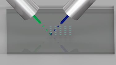
When we think of silk, our minds often turn to its soft, luxurious feel in textiles. But silk is far more versatile. It is a remarkable material with properties that extend beyond fabrics and into engineering and biomedical applications. My recent research focuses on 3D-printed silk-based materials, created using reactive inkjet printing. These materials boast a unique combination of rigidity and flexibility, a mix of mechanical properties that can be adjusted for diverse applications. We’ve even used silk to encapsulate proteins, which adds stability and enhances functionality.
However, one mystery remains: why do these materials exhibit such extraordinary properties? To address this, we turned to advanced imaging technologies.
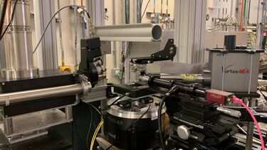
Last month, I had the privilege of conducting experiments at the I13-1 Coherence Beamline at Diamond Light Source, a world-class synchrotron facility. This was not my first experience at Diamond. On a previous visit, I worked alongside Nicholas T. H. Farr from the School of Chemical, Materials and Biological Engineering and Professor John Rodenburg, an eminent researcher in X-ray ptychography. During that initial trip, I was introduced to ptychography, a cutting-edge imaging technique that uses coherent X-rays to generate highly detailed 3D reconstructions of materials.
Encouraged by the insights gained during that collaboration, I applied for beamtime to explore how these imaging methods could shed light on my own work with silk-based 3D-printed biomaterials.
X-ray ptychography offers a non-destructive way to peer inside materials at the nanoscale, revealing details that traditional imaging techniques might miss. By combining coherent X-rays with advanced computational methods, it creates detailed three-dimensional images of internal structures.
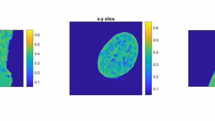
Using this technique, we aim to uncover the structural and molecular reasons behind the unique properties of silk-based materials. Specifically, we’re examining how reactive inkjet printing influences the interplay of rigidity and flexibility. Additionally, we’re investigating how encapsulated proteins interact with the silk matrix to stabilise and enhance the material’s functionality and influence the reactive printing process.
Understanding the properties of these materials could lead to transformative applications, particularly for biomedical applications and in biosensing. Silk-based materials have the potential to be used in sensors capable of detecting toxins, pathogens, and other hazardous substances as well as for rapid disease diagnostics. Their adaptability makes them promising candidates for responsive, biodegradable devices, bridging the gap between fundamental science and real-world needs.
This research would not have been possible without the support and expertise of a fantastic team. I am especially grateful to Nicholas T. H. Farr, Mahendra P. Raut, Leo Fish, Jonathan Hinchliffe, and Professor Chris Holland for their contributions. Special thanks also go to beamline scientists Zifan Wang and Darren Batey for their guidance and assistance throughout the measurements. Finally, I must acknowledge Professor John Rodenburg, whose insights into X-ray ptychography were invaluable.

Future perspectives
As we begin analysing the data collected, I am excited to uncover new insights into the behaviour of silk-based materials. This is just the beginning of a journey to understand their extraordinary potential. By combining innovative 3D printing techniques with advanced imaging, we hope to answer fundamental questions and pave the way for technologies that can address critical challenges in biosensing and beyond.
Stay tuned for updates as we dive deeper into the data and explore the exciting possibilities of this work! Please find my contact details on my staff profile page, and
Acknowledgements – MG38437-1, School of CMBE, SM3 ()
This article was written by Dr David A Gregory.